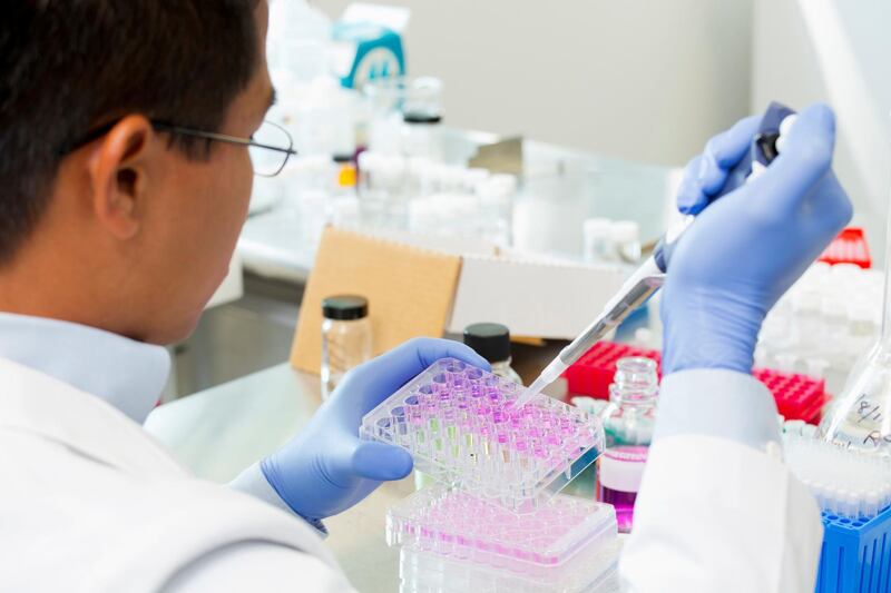

Researchers in Abu Dhabi have developed a new paper-based method to create and preserve small lab-grown tumours, on which anti-cancer drugs can be tested.
Scientists are likely to get more accurate results when they try drugs out on 3D tumours, compared to other commonly used methods, researchers at New York University Abu Dhabi said.
While small 3D tumours are already being grown and used in laboratory tests, this new technique offers an improved way to create them in large numbers and store them at low temperatures for future use, researchers said.
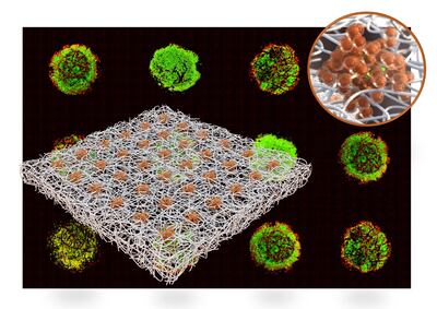
The technique involves growing 3D cancer tumours, that can be as little as 0.1 millimetres in diameter, on filter paper – similar to coffee filters.
The tumours can be produced in large numbers at one location and stored, before being shipped out to laboratories and used for drug tests at a later date.
"You can roll the paper and put it in liquid nitrogen and store it for the future," said Mohammad Qasaimeh, an NYUAD assistant professor of mechanical and biomedical engineering, in whose laboratory the work was done.
"When you want to test your drug, you retrieve these papers, these 3D arrays."
Currently, potential anti-cancer drugs are often tested on 2D sheets of cells, but these often give unreliable results.
In their paper in the scientific journal Lab on a Chip, the NYUAD team described such sheets of cells as "overly simplified".
“Eighty per cent of candidate drugs will fail. They will show positive effects on 2D cells, but fail with animal tests or clinical trials,” Mr Qasaimeh said.
Mr Qaisameh said 3D tumours were increasingly being used instead of 2D sheets of cells and in some instances, instead of animal tests.
“Although animal models may give insight into key biological responses, the anatomy and physiology of animals are profoundly different from those of humans, and there are ethical concerns regarding their use in research,” the researchers wrote.
Their technique, described in a paper titled, Cryopreservable Arrays of Paper-based 3D Tumour Models for High Throughput Drug Screening, uses tiny "microspots" on the paper.
Small aggregations of cancer cells are able to form on these microspots.
To see how effective their method was, the researchers grew 3D breast cancer tumours and tested out a widely used cancer chemotherapy drug, cisplatin.
Tests using the 3D tumours accurately indicated how well the drug worked.
Paper has emerged as an "attractive, simple tool" for growing 3D cell cultures, helped by the nature of cellulose – the main structural chemical of plant cell walls.
Cellulose fibres create an appropriate “niche” for 3D groups of cells to grow.

Until now, however, there has also been a lack of “off-the-shelf” paper-based methods to grow and preserve small 3D tumours.
Their methods, the NYUAD team said, involved simple, scalable and inexpensive processes.
The researchers said their method could also be applied to personalised or precision medicine, which tailors treatment to the genetic characteristics of a patient’s tumour.
A key emerging way to treat cancer, this improves the effectiveness of treatments and reduces side effects.
Other researchers behind the new study include Bisan Samara, a former research assistant in Dr Qasaimeh’s laboratory and the first author of the paper.