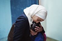
ABU DHABI // Doctors have been issued with guidelines aimed at the earlier detection and more effective treatment of breast cancer.
Issued by the Health Authority - Abu Dhabi (Haad) in collaboration with the Tawam Molecular Imaging Centre (TMIC), the guidelines specify treatments based on individual diagnoses.
They incorporate international standards and instructions adapted for patient requirements.
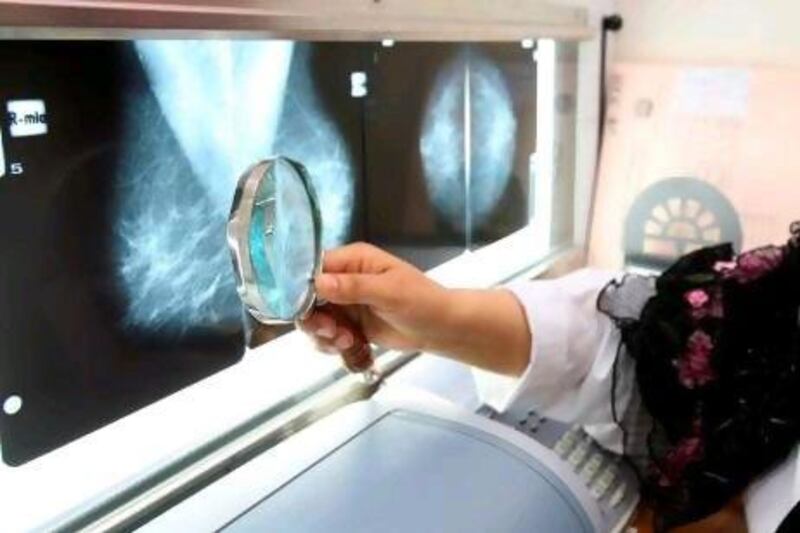
The most influential part of the new guidelines is recognising positron emission tomography (Pet) scans as a standard requirement for imaging once a diagnosis is made, said Dr Jala Taher, the section head of cancer control and prevention at Haad.
The imaging technology can better detect the spread of cancer than traditional forms of imaging.
The guidelines are a response to the more aggressive forms of breast cancer that are appearing in younger patients, Dr Taher said, adding that nearly half of the cases diagnosed in Abu Dhabi were in women under the age of 46.
"We shouldn't just follow what others are implementing," she said. "We need to customise things to our situation."
Figures from Haad show breast cancer is the most common cancer in the UAE, accounting for 25 per cent of all cases and 45 per cent of female cases in Abu Dhabi in 2010.
On average, 170 women are diagnosed with breast cancer each year in the emirate. It is the second-leading cause of death for women after cardiovascular disease.
Dr Taher said the level of awareness had noticeably increased.
"Now, more women go for regular screening than ever before," she said. "As a result, the prevalence of late-stage presentation of breast cancer reduced dramatically, from 65 per cent in 2007 to 25 per cent in 2010."
Haad is holding workshops to inform doctors on the new guidelines and educate them on how and when to use the technology.
The instructions are the first of a series of new cancer-management guidelines to be issued by Haad and TMIC. Guidelines for the management of cervical cancer and colorectal cancer will follow.
Previously, doctors were following international guidelines , typically those set by the National Comprehensive Cancer Network in the US, which do not recommend the use of Pet scans, opting instead for conventional imaging methods.
But these technologies only capture the structure of a mass, whereas functional imaging such as that of a Pet scan identifies the cancer's active areas and its spread to other parts of the body.
The most common solution used as a radiotracer in Pet scans for cancer is glucose, as cancer cells consume more glucose than normal cells. It is injected into the body, then picked up by the scan.
The cost of a Pet scan is between Dh8,000 and Dh14,000 for a single imaging session. But TMIC works with major insurance companies, including Daman and Abu Dhabi National Insurance Company, to ensure coverage for its patients.
Other insurance companies are also reimbursing patients.
With rigorous screening programmes introduced in the US for nearly three decades, Pet scans are not considered a cost-efficient method for managing the disease.
In the UAE, however, doctors are treating women in their 20s with advanced stages of the disease, said Dr Muhammad Chaudhry, the medical director of TMIC.
"The last time in the US they saw a 5cm mass on a 25-year-old female was about 30 years ago," Dr Chaudhry said. "So yes, you have to modify your local treatment."
Another benefit of Pet scans is in monitoring treatment results, he said.
"Initially when you're giving chemotherapy … it would start killing cells and the tumour would begin to shrink," Dr Chaudhry said. "But most treatments now stop the cell from growing, so the mass will look the same to you if you continue to use structural imaging.
"If you use functional imaging, however, you would see the tumours are not metabolically active any more and you would know that the treatment you gave is effective."
Dr Taher said mammograms were not an effective option for young women because the imaging device detected the contrast between breast tissue and cancer cells using shades of black, white and grey.
"The older the female, the less breast tissue she will have," she said. "All will be replaced by fat so there will be good contrast visible.
"Whereas in a young woman, who has a lot of breast tissue, all you will see is white. If there's any cancer it will be hidden behind this white."
Doctors advise girls with a family history of breast cancer to have an MRI scan.
Experts hope the guidelines will ensure timely, quality management of the disease, increasing the survival rate.
"This method could help detect metastasis that could have been missed using previous methods," Dr Taher said. "That means earlier and more effective treatment, and saving a patient's life."